
A typical radiologist is under extreme pressure and time constraints to perform accurate readings. On average, a radiologist is afforded seconds to interpret each image in a series to meet the workload demands in an 8-hour workday. The need to interpret more images in less time continues to increase with advancements in imaging modalities and pulse sequences creating additional pressure and demand. The workload dynamic on radiologists at large medical facilities has become untenable. In some countries, such as China, there is only one radiologist for every 40,000 people.
In many cases, such as diagnostic imaging in individuals presenting with a probable stroke, patient outcomes are directly correlated with speed and the care dynamic is extremely time sensitive. Thus, the ability for radiologists to know which patient to examine first can improve patient outcomes. Furthermore, imaging modalities such as Magnetic Resonance Imaging (MRI) take a long time in some instances to acquire a series of images. These areas are all ripe for leveraging the use of artificial intelligence (AI) and machine learning (ML) to speed up medical imaging diagnosis.
Reducing Noise
AI has been utilized to reduce the time it takes for an MRI scan from one hour to as little as 15 minutes. Only a portion of the normal data for an MRI scan is collected. Deep learning-based methods reform image reconstruction into a data-driven supervised learning problem of finding a mapping between the sensor and the image domain. In this way, artificial intelligence is used to fill in the gaps and accurately complete the image. Additionally, AI reduces the “noise” in the image, providing a clearer picture than traditional MRI. AI can also be used to aid in making MRI scanners more intelligent with machine-controlled choices of protocols and calibrations, selection of optimal imaging techniques, and adaptation to individual variations in anatomy and patient. This results in image acquisitions made faster, with more reliable reconstructions, fewer artifacts and reduced noise. This will allow for more patients to receive an MRI scan from the same MRI machine in one day and for a scan to be more affordable for patients. Furthermore, this could lead to more use of MRI in more urgent situations where Computerized Tomography (CT) scans today may be typically preferred.

Workflow Improvement
AI can help radiologists more intelligently prioritize their work. AI algorithms can be used to help identify critical images of patients to interpret first. In this way AI can assist in prioritizing the radiologist’s workflow and moving the most critical images to the top of a worklist. Natural language processing can also be utilized to provide insights of data to improve workflows. Workflow improvement can lead to the patients requiring the most urgent care receiving it first, and thus improve patient outcomes.
Image Interpretation
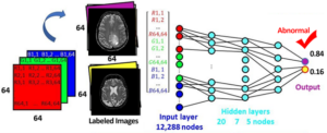
AI can also help radiologists with image interpretation. Machine learning tools can be leveraged to provide another diagnostic opinion about the image, such as if an image has any abnormality present. A deep learning neural network can be trained using labeled images of diseases and normal conditions present. The labeled 2D images in matrix form can be input to the deep learning model as a 1D vector and numerous hidden layers can be added to the neural network to help prevent overfitting. After training has occurred and new image is input to the trained neural network, by looking at the output insights can be gleaned. In this example, an output greater than 0.5 indicates an abnormality present and an output less than or equal to 0.5 indicates normal conditions. By providing radiologists with an opinion before they look a series of images, they can more quickly arrive at their own diagnostic opinions and move through images faster.
Computer vision and machine learning can also be utilized to automate image segmentation tasks such as the segmentation of organs and lesions images. These tasks can help reduce the amount of time spent looking at innocuous details and bring suspicious abnormalities of greater interest to the forefront.
Utilizing AI as a triage mechanism and in support of more efficient workflows hold great promise in the domain of medical imaging diagnosis. It is plausible that every image will one day be pre-screened and analyzed by a computer prior to human interaction, providing valuable insight and maximizing the efficiency of radiologists and medical imaging professionals. For medical imaging, the diagnostic challenge is to move beyond screening test sets and apply the power of algorithms to consider variations in patient characteristics, disease incidence, and severity. The culmination of these efforts will improve resource utilization, accelerate accurate diagnosis, and ultimately drive optimal clinical outcomes.
If you’re ready to explore the potential of AI to solve your organization’s medical imaging research needs, Ellumen’s AI experts are here to help. Keep an eye out for the next part of Ellumen’s blog series exploring topics related to AI innovation in medical imaging.
Meet Ellumen’s AI Experts
Are you looking to utilize AI for your next technology or integrate AI into your current systems? Feel free to reach out to us.

Iyanuoluwa Odebode, Ph.D. is Ellumen’s artificial intelligence/machine learning expert, supporting the Innovation Lab project by identifying and researching new & emerging technology. He also serves as an adjunct professor in cybersecurity at University of Maryland Baltimore County. Prior to his work at Ellumen, Iyanuoluwa built an algorithm for repurposing old drugs for new use using the DReiM methodology. He has been published in ResearchGate and IEEE for his research on machine learning. Iyanuoluwa completed his master’s in bioinformatics at Morgan State University and his Ph.D. in information systems (machine learning/AI) at University of Maryland Baltimore County.

Todd R. McCollough is a Software Engineer for Ellumen. He works primarily with the CVIX/VIX Services which support image viewers and applications to provide the ability for users to query, retrieve and manipulate VA and DoD medical images and image artifacts. Todd is a co-inventor on several issued U.S. patents and is passionate about discovering novel ways to image patients and improve patient care. Todd received his bachelor’s and master’s degrees in biomedical engineering from Northwestern University.